Search Results for author:
Found 6 papers, 1 papers with code
DEMIST: A deep-learning-based task-specific denoising approach for myocardial perfusion SPECT
There is an important need for methods to process myocardial perfusion imaging (MPI) SPECT images acquired at lower radiation dose and/or acquisition time such that the processed images improve observer performance on the clinical task of detecting perfusion defects.

Need for Objective Task-based Evaluation of Deep Learning-Based Denoising Methods: A Study in the Context of Myocardial Perfusion SPECT
Our objectives were to (1) investigate whether evaluation with these FoMs is consistent with objective clinical-task-based evaluation; (2) provide a theoretical analysis for determining the impact of denoising on signal-detection tasks; (3) demonstrate the utility of virtual clinical trials (VCTs) to evaluate DL-based methods.

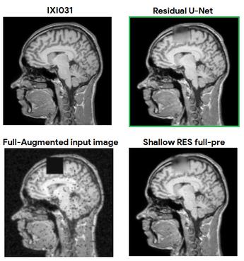
Synthetic PET via Domain Translation of 3D MRI
Historically, patient datasets have been used to develop and validate various reconstruction algorithms for PET/MRI and PET/CT.
A physics and learning-based transmission-less attenuation compensation method for SPECT
The proposed method uses data acquired in the scatter window to reconstruct an initial estimate of the attenuation map using a physics-based approach.
Medical Physics
Observer study-based evaluation of a stochastic and physics-based method to generate oncological PET images
In this study, we develop a stochastic and physics-based method to generate realistic oncological two-dimensional (2-D) PET images, where the ground-truth tumor properties are known.
Medical Physics Image and Video Processing
A Bayesian approach to tissue-fraction estimation for oncological PET segmentation
Conventional segmentation methods are typically designed to assign each voxel in the image as belonging to a certain tissue class.

