Search Results for author:
Found 10 papers, 6 papers with code
Registration by Regression (RbR): a framework for interpretable and flexible atlas registration
In human neuroimaging studies, atlas registration enables mapping MRI scans to a common coordinate frame, which is necessary to aggregate data from multiple subjects.
P-Count: Persistence-based Counting of White Matter Hyperintensities in Brain MRI
White matter hyperintensities (WMH) are a hallmark of cerebrovascular disease and multiple sclerosis.


PEPSI: Pathology-Enhanced Pulse-Sequence-Invariant Representations for Brain MRI
Remarkable progress has been made by data-driven machine-learning methods in the analysis of MRI scans.

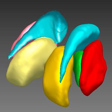
H-SynEx: Using synthetic images and ultra-high resolution ex vivo MRI for hypothalamus subregion segmentation
Materials and Methods: We trained our deep learning method, H-synEx, with synthetic images derived from label maps built from ultra-high resolution ex vivo MRI scans, which enables finer-grained manual segmentation when compared with 1mm isometric in vivo images.
Quantifying white matter hyperintensity and brain volumes in heterogeneous clinical and low-field portable MRI
Brain atrophy and white matter hyperintensity (WMH) are critical neuroimaging features for ascertaining brain injury in cerebrovascular disease and multiple sclerosis.
Brain-ID: Learning Contrast-agnostic Anatomical Representations for Brain Imaging
We present new metrics to validate the intra- and inter-subject robustness of Brain-ID features, and evaluate their performance on four downstream applications, covering contrast-independent (anatomy reconstruction/contrast synthesis, brain segmentation), and contrast-dependent (super-resolution, bias field estimation) tasks.
A Lightweight Causal Model for Interpretable Subject-level Prediction
Recent years have seen a growing interest in methods for predicting a variable of interest, such as a subject's diagnosis, from medical images.
SynthSeg: Segmentation of brain MRI scans of any contrast and resolution without retraining
Here we introduce SynthSeg, the first segmentation CNN robust against changes in contrast and resolution.


A Contrast-Adaptive Method for Simultaneous Whole-Brain and Lesion Segmentation in Multiple Sclerosis
Here we present a method for the simultaneous segmentation of white matter lesions and normal-appearing neuroanatomical structures from multi-contrast brain MRI scans of multiple sclerosis patients.

A Modality-Adaptive Method for Segmenting Brain Tumors and Organs-at-Risk in Radiation Therapy Planning
In this paper we present a method for simultaneously segmenting brain tumors and an extensive set of organs-at-risk for radiation therapy planning of glioblastomas.
