Computed Tomography (CT)
294 papers with code • 0 benchmarks • 14 datasets
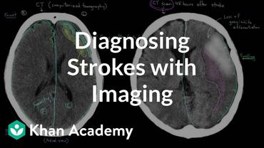
The term “computed tomography”, or CT, refers to a computerized x-ray imaging procedure in which a narrow beam of x-rays is aimed at a patient and quickly rotated around the body, producing signals that are processed by the machine's computer to generate cross-sectional images—or “slices”—of the body.
( Image credit: Liver Lesion Detection from Weakly-labeled Multi-phase CT Volumes with a Grouped Single Shot MultiBox Detector )
Benchmarks
These leaderboards are used to track progress in Computed Tomography (CT)
Libraries
Use these libraries to find Computed Tomography (CT) models and implementationsDatasets
Most implemented papers
COVID-CT-Dataset: A CT Scan Dataset about COVID-19
Using this dataset, we develop diagnosis methods based on multi-task learning and self-supervised learning, that achieve an F1 of 0. 90, an AUC of 0. 98, and an accuracy of 0. 89.
MULAN: Multitask Universal Lesion Analysis Network for Joint Lesion Detection, Tagging, and Segmentation
When reading medical images such as a computed tomography (CT) scan, radiologists generally search across the image to find lesions, characterize and measure them, and then describe them in the radiological report.
UNet++: Redesigning Skip Connections to Exploit Multiscale Features in Image Segmentation
The state-of-the-art models for medical image segmentation are variants of U-Net and fully convolutional networks (FCN).
Evaluate the Malignancy of Pulmonary Nodules Using the 3D Deep Leaky Noisy-or Network
The model consists of two modules.
The KiTS19 Challenge Data: 300 Kidney Tumor Cases with Clinical Context, CT Semantic Segmentations, and Surgical Outcomes
The morphometry of a kidney tumor revealed by contrast-enhanced Computed Tomography (CT) imaging is an important factor in clinical decision making surrounding the lesion's diagnosis and treatment.
The Liver Tumor Segmentation Benchmark (LiTS)
In this work, we report the set-up and results of the Liver Tumor Segmentation Benchmark (LiTS), which was organized in conjunction with the IEEE International Symposium on Biomedical Imaging (ISBI) 2017 and the International Conferences on Medical Image Computing and Computer-Assisted Intervention (MICCAI) 2017 and 2018.
3D Context Enhanced Region-based Convolutional Neural Network for End-to-End Lesion Detection
3D context is known to be helpful in this differentiation task.
Disentangled Representation Learning in Cardiac Image Analysis
We can venture further and consider that a medical image naturally factors into some spatial factors depicting anatomy and factors that denote the imaging characteristics.
Framing U-Net via Deep Convolutional Framelets: Application to Sparse-view CT
X-ray computed tomography (CT) using sparse projection views is a recent approach to reduce the radiation dose.
Generative Adversarial Networks for Image-to-Image Translation on Multi-Contrast MR Images - A Comparison of CycleGAN and UNIT
Here, we evaluate two unsupervised GAN models (CycleGAN and UNIT) for image-to-image translation of T1- and T2-weighted MR images, by comparing generated synthetic MR images to ground truth images.

 LUNA
LUNA
 PadChest
 LUNA16
 ChestX-ray8
 LiTS17
 COVID-CT
 MosMedData
 BIMCV COVID-19
 CC-19
CC-19
