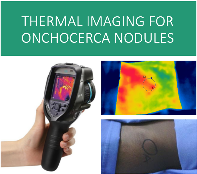
Evaluation of Non-Invasive Thermal Imaging for detection of Viability of Onchocerciasis worms
Onchocerciasis is causing blindness in over half a million people in the world today. Drug development for the disease is crippled as there is no way of measuring effectiveness of the drug without an invasive procedure. Drug efficacy measurement through assessment of viability of onchocerca worms requires the patients to undergo nodulectomy which is invasive, expensive, time-consuming, skill-dependent, infrastructure dependent and lengthy process. In this paper, we discuss the first-ever study that proposes use of machine learning over thermal imaging to non-invasively and accurately predict the viability of worms. The key contributions of the paper are (i) a unique thermal imaging protocol along with pre-processing steps such as alignment, registration and segmentation to extract interpretable features (ii) extraction of relevant semantic features (iii) development of accurate classifiers for detecting the existence of viable worms in a nodule. When tested on a prospective test data of 30 participants with 48 palpable nodules, we achieved an Area Under the Curve (AUC) of 0.85.
PDF AbstractTasks
Datasets
Introduced in the Paper:
Niramai Oncho Dataset