GlaS (Gland Segmentation in Colon Histology Images Challenge)
Introduced by Sirinukunwattana et al. in Gland Segmentation in Colon Histology Images: The GlaS Challenge Contest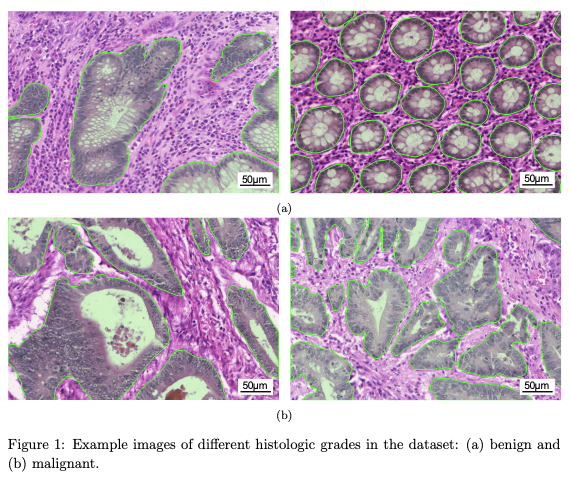
The dataset used in this challenge consists of 165 images derived from 16 H&E stained histological sections of stage T3 or T42 colorectal adenocarcinoma. Each section belongs to a different patient, and sections were processed in the laboratory on different occasions. Thus, the dataset exhibits high inter-subject variability in both stain distribution and tissue architecture. The digitization of these histological sections into whole-slide images (WSIs) was accomplished using a Zeiss MIRAX MIDI Slide Scanner with a pixel resolution of 0.465µm.
Source: Sirinukunwattana et al.
Papers
| Paper | Code | Results | Date | Stars |
|---|


